Лечение в Германии Center for medical treatment in Germany

Tel.: +49 89 1500 15 45 E-Mail: registratura@ger-doc.de
en  de  ru 

Tag: cancer germany

Multiple adenocarcinoma

Diagnosis and treatment in Munich

Diagnosis and treatment of oncological diseases in Munich

Question:

Please consider the possibility of treatment / extending the life of comfort for my brother.

Oncology Clinic in Belarus diagnoses (by a consultation) Generalized form of cancer (adenocarcinoma) with no established primary focus with the defeat of the skeleton and bone marrow, the development of DIC.

Diagnosis: Secondary malignant neoplasm of bone and bone marrow (S79.5).

Multiple mts adenocarcinoma in bones of the skeleton of an unknown primary tumor. State after trephine biopsy of the tumor left iliac region (22.03.2013g.). Chronic DIC. Progressive liver-kidney failure.

TNM: Tx Nx M1 stage IV

These morphological studies:

Adenocarcinoma NOS [8140/3]

cell tumor (most likely adenocarcinoma)

Metastases: regional – not found

remote – in the bones

19/03/2013 Analyses:

Blood for tumor markers: CEA> 1000 ng / ml, CA19-9> 1000 kg / ml ACE 2,08 ME / ml PSA total. 0.55 ng / ml

Coagulation:

APTT: 32.3 sec

Fibrinogen 1.6 g / l

Thrombin time: 21.2 seconds

Large bruises all over his body.

In its current state: morphine analgesia 4 p / day. In the mind.

Please advise whether it is possible hospitalization and emergency treatment options for the patient.

P.S. There is a history of the disease in the pdf in Russian.


Answare:

We are ready at any time convenient for you to organize a consultation with our professors and if necessary treatment at the clinic.
To concretize the treatment of adenocarcinoma of the proposals, please provide us with the following information:
- A copy of the first page of your passport (for authorization).
- Case history and all the results of this research, etc.
- The date of the desired arrival.

Payment for medical care you will make doctors and hospitals directly.


Diagnosis of testicular seminoma
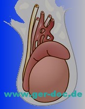
Treatment of seminoma in Munich

Seminom

Question
Recently began to notice a gradual painless enlargement and discomfort in the right half of the scrotum. addressed to the urologist, performed ultrasound, and the appendage of the left testicle is not changed. The right testicle with fuzzy rough contours, heterogeneous parenchyma due to inclusions hypoechoic nodular structure, the size of eggs 11.2 x6, 7 cm The epididymis is not changed. When DRC is marked diffuse increase in blood flow in the right testicle.
AFP – 1.0 IU / mL hCG – 1.1 mIU / mL LDH – 2575 units / l.
Our doctors suspect cancer.
What is required for the passage of diagnosis in Munich?

Answer:
To assist in the organization of diagnosis and treatment seminoma testicular (ie, testicular cancer) you want to start to send the case history.

Mammary cancer

Mammary cancer

Mammary cancer

Mammography

Examination was accomplished in standard projections: straight and oblique. The mammary glands are symmetrical.
The areolas are output at a contour. The skin is not thickened, not condensed. The bands of fibrous tissue – Cooper’s ligaments are left from the skin. The premammary space is not constricted. A tissue of the mammary gland is presented in central and lateral areas by the glandular tissue, in medial by adipose and line diffuse fibrosis in all quadrants. The density of both breasts has the 2nd grade.
At this background of pathological masses, no micro calcinations are detected
Single micro calcifications are visualized on the border of inferior quadrants of the left breast.
A vascular grid is well defined.
No abnormalities of the lymph flow zones on the right and on the left.

ECHO MAMMOGRAPHY

The mammary glands are symmetrical. The skin is not changed.
The mammary glands have the usual structure for this age.
The differentiation of tissues is evident sufficiently.
Cooper’s ligaments which go deep are thickened and condensed.
Galactophorous ducts are moderately dilated till 2.0-2.5 mm.
No pathological parts of vascularization are detected.
Parts near areola are without any changes.

Impression: No signs of malignizalion are detected. ACR1 is for the right and ACR2 is for the left breast. The density of both breasts has the 2nd grade. Ultrasonic signs of diffuse changes of the breasts by the type glandular mastopathy.

General examination: The patient complaints for having a mass in the upper-internal quadrant of the left breast, which she noted in August 2012. She has felt enlargement of this mass in sizes from August till today.
Data of previous consultations – 28 of May 2012 – mammography, Breast use – ACR1 is on the right, ACR2 is on the left.
For the period from May till November felt changes in hormone therapy, climate changes (lived in Uzbekistan in summer).
St. localis: The mammary glands are symmetrical, and the regular color. The skin is not condensed under them. A mass of rounded form with solid non-crushing consistency and unconsolidated with surrounding tissues till 2 sm is palpated in the upper-internal quadrant of the left breast. Palpation is without pain. The axillary lymph nodes are not palpated.
Breast ultrasound: The mammary glands are placed typically; the skin is without any changes. The differentiation of tissues is expressed in both breasts. The correlation of adipose and glandular tissue: 1/3.
The Galactophorous ducts are thickened a little in both breasts till 2.0-2.1 mm. Cooper’s ligaments are not thickened and not expanded.
A dimensional, massive, hypo echoic and avascular mass of oval form 17.4×10.3 mm, which has calcinations in the center is located in the left breast in the upper-internal quadrant. No areas of pathological vascularizations. The regionary lymph nodes are not increased.

Impression: Ultrasonic signs of moderate diffuse changes of both breasts by the type of glandular mastopathy. Signs of dimensional mass of the left breast (fibro adenoma?). Biopsy of the pathological mass of the left breast is recommended.

HYSTOLOGY EXAMINATION

Tissue labeling, number of objects: №1 – biopsy of the tumor of the left breast.
Clinical diagnosis: Malignized tumor of the left breast.
Macroscopic description: 4308-01, 02 – two (2) grey color bands with brown areas and with the length of 1.7 sm each.
Microscopic description: Conclusion: Enlargements of invasive ductal carcinoma Grade III are revealed in all biopsy samples in the tissue of the mammary gland.

Mammography

Examination was accomplished in oblique views. The mammary glands are symmetrical and not deformed. The skin is not thickened, not condensed.
The bands of fibrous tissue – Cooper’s ligaments are left from the skin. The premammary space is not constricted.
A tissue of the mammary gland is mainly presented by glandular tissue, in medial areas by adipose tissue and line diffuse fibrosis in all quadrants.
High intensity without clear contours of rounded mass till 18 mm in diameter, which is located retromammary, containing single polymorph micro calcinations is revealed at this background on the border of internal quadrant of the left breast. Behind the mass, a single line calcination is visualized. Abnormality of architectonic of structured picture of the central part of the left breast due to stroma deformation is revealed. A vascular grid is well defined.
No abnormalities of the lymphatic flow on the left and on the right.
IMPRESSION: ACR1 is for the right and ACR4 is for the left breast. X-ray signs of dimensional mass of the left breast.

MAGNETIC RESONANCE IMAGING BREASTS

Description of the research:
A mass of rounded form with even contours and sizes 16x11x17 mm due to active accumulation of contrast material more on the periphery with washing-out to the 3rd minute is revealed in the left breast in the deep areas of the breast in the upper-internal quadrant. The mass is surrounded by the rich vascular grid.
No pathological changes in the right breast. No areas of accumulation of contrast material in the lymphatic nodes are detected.
No bone destructive changes.
IMPRESSION: MR picture of the left breast damage, probably, having neoplastic nature. Biopsy is recommended to make verifications.
Magnetic resonance tomograph EXCELART Vantage Atlas-X 1,5 T Toshiba

Complaints: for having occasional pain in the breasts and having a tumor-like mass of the left breast.
History of the disease: After self-palpation in April 2012, the patient noted a tumor-like mass for the first time.
General examination: Micromastia. The breasts are asymmetrical in the inferior-internal quadrant of the left breast. Vertically oriented tumor-like mass of the solid consistency with sizes 20-27 mm is palpated (the patient notes enlargement of the tumor after biopsy). No skin symptoms. Regionary lymph nodes are palpated not clear. No nipple discharge.
Diagnosis:
C50.3 Cancer of the left breast T3NxMx
Differentiated diagnosis: diagnosis is verified. Plan of examination: Mammography.
Treatment: Additional consultation of oncomammologist and chemotherapist for defining indications to have possible neoajuvant chemotherapy.

Рecommendation

Treatment in Germany.
Ideal: treatment in Munich.

Cancer treatment with Cyberknife©

Cancer treatment in Germany

Cancer treatment in Germany with Cyberknife

Cancer treatment in Germany with Cyberknife©


“Proton therapy” and Cyberknife© are the most advanced high-tech method in the treatment of cancer with radiation therapy.

In the radiology clinic of Munich cancer patients are given the opportunity to be treated by one of the most modern methods of treatment of cancer. Them is a highly accurate, robot-assisted and high-tech method of radiation known as Cyberknife©.

This method has successfully treated more than 10,000 patients from all over the world in the Specialized Clinic Munich Cyberknife© are in Germany.

Therapy by the Cyberknife© is not only the most gentle, and painless for the patient, and always on an outpatient basis. Producing point exposure through special terminals and the concentration of rays that are at the intersection have significant energy potential. Thus, no interference with a surgical scalpel and cancerous tissue is destroyed, without exposing the ravages of its surrounding healthy tissue. No longer requires a painful and difficult for the body to open surgery. Due to this method, doctors can avoid possible complications of anesthesia, a multi-day hospital stay, postoperative treatment costly and lengthy recovery period of the patient.

A particular advantage of the approach in Cyberknife© is an painless, but very effective treatment of a number of cancers, for example: the spine cancers, spinal tumors, brain metastases and brain cancer, lung cancersand

. Through the use of several terminals, this approach makes it possible to divide a strong dose for a few much smaller exposures, each of which by itself is completely harmless, and at the intersection of rays created the necessary concentration. Thus, it was possible to conduct therapy in the most sensitive places and bodies, and the surrounding healthy tissue is not damaged.

Outpatient treatment Cyberknife© is held only once and does not require any restrictions during the daily life and the usual routine patient remains Following this therapy session.

Due to the strong, but the point dose therapy Cyberknife© is usually a one-time procedure and lasts depending on the requirements of a few minutes. German experts recommend the checks no later than one half of the year.

For more information about Cancer treatment with Cyberknife© please go to medical advice.