|
|
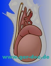
Category Archives: Оncology
 gastric cancer Patient:
A female friend of mine ( aged 25 ) from Dubai was diagnoses as having gastric cancer 2 months ago. She has had part of her stomach removed in a hospital in Shiraz ( Iran ). She has been told by the doctor who did the operation, to go immediately to Germany for the continuation of her treatment. Please kindly advise as to exactly where in Germany is best for her to go for the continuation of her treatment.
Thanking you for your kind attention, I look forward to hearing from you soon.
Answer:
Thank you for contacting our Medical Services for information on obtaining medical care for you.
In order for our physicians to properly review this case and determine an appropriate course of treatment, we would need additional information.
Please complete the attached New Patient Form and sign the Financial Policy statement and return the forms along with all pertinent medical records in German or English (including any CT scans, MRIs or x-rays, etc).
In order to give a 2nd opinion the doctors need to know/receive:
- Clinical notes, medical history,
- Treatment he has received up till now Chemo? Dosage & frequency?
- Radiation? Dosage & frequency?
- His symptoms?
- Current list of medication
- List of operations/surgeriesOperative report
- Pathology slides and reports.
- A copy of the images of his PET CT Scan on a CD sent to our address below.
Also, as of January 1, 2000 we are required to verify patient identity. Please review the attached document which list acceptable forms of identification.
Please send a copy of his photo identification to our Medical Services Department with his completed patient forms and medical records and be prepared to show his original photo documentation when he arrives for his appointment(s).
You may return the forms/medical records via e-mail and you can return these forms to our office via regular mail, DHL, Fed Ex or UPS to our address.
Once we have received this information, our physicians will review the case and advise accordingly. Please be advised that there may be a fee associated with this consult.
 Diagnosis and treatment of oncological diseases in Munich Question:
Please consider the possibility of treatment / extending the life of comfort for my brother.
Oncology Clinic in Belarus diagnoses (by a consultation) Generalized form of cancer (adenocarcinoma) with no established primary focus with the defeat of the skeleton and bone marrow, the development of DIC.
Diagnosis: Secondary malignant neoplasm of bone and bone marrow (S79.5).
Multiple mts adenocarcinoma in bones of the skeleton of an unknown primary tumor. State after trephine biopsy of the tumor left iliac region (22.03.2013g.). Chronic DIC. Progressive liver-kidney failure.
TNM: Tx Nx M1 stage IV
These morphological studies:
Adenocarcinoma NOS [8140/3]
cell tumor (most likely adenocarcinoma)
Metastases: regional – not found
remote – in the bones
19/03/2013 Analyses:
Blood for tumor markers: CEA> 1000 ng / ml, CA19-9> 1000 kg / ml ACE 2,08 ME / ml PSA total. 0.55 ng / ml
Coagulation:
APTT: 32.3 sec
Fibrinogen 1.6 g / l
Thrombin time: 21.2 seconds
Large bruises all over his body.
In its current state: morphine analgesia 4 p / day. In the mind.
Please advise whether it is possible hospitalization and emergency treatment options for the patient.
P.S. There is a history of the disease in the pdf in Russian.
Answare:
We are ready at any time convenient for you to organize a consultation with our professors and if necessary treatment at the clinic.
To concretize the treatment of adenocarcinoma of the proposals, please provide us with the following information:
- A copy of the first page of your passport (for authorization).
- Case history and all the results of this research, etc.
- The date of the desired arrival.
Payment for medical care you will make doctors and hospitals directly.
Diagnosis and treatment of cancer in Munich LIVER: Location – in the right upper quadrant.
Form-variant of the norm.
The contours are smooth, clear.
The capsule is not changed.
Dimensions are not changed: the right lobe of the thickness of 129 mm (normal, 120-125 mm), left lobe – the thickness of 76 mm (normal 50-60 mm)
The structure of the parenchyma diffuse-not homogeneous.
Echogenicity, increased vascular drawing is saved.
Gate Vienna anehogennoe visualized as a tubular structure is not expanded, with a diameter of 10 mm (normal up to 13 mm). The hepatic veins are dilated. Lower hollow Vienna diameter of 19 mm. Choledoch – 5 mm (normal up to 6 mm) at the gate of the liver. Intrahepatic bile ducts are not dilated Volumetric structures have been identified.
Recommendation: run diagnostics in Germany.
GALLBLADDER: Location – the usual.
The form has not changed. The contours are smooth, clear.
Dimensions are not changed: dlinnik-78 mm (normal range 60-100 mm) diameter, 22 mm (standard 30 mm), cavity – free, anehogennoe content.
Stones, structures have been identified.
Not thickened walls (norm to 3 mm).
Echogenicity of the wall – the average.
The structure of the wall – homogeneous.
Choledoch not extended, pass all over.
Recommendation: spend a treatment in Germany.
Pancreas: Location typical. The contours are smooth, clear.
Form-variant of the norm.
Dimensions are not changed: a head-23mm (normal up to 11-30 mm), the body is 18 mm (normal 4-21 mm), tail 22 mm (normal, 7-28 mm).
The structure of the parenchyma – diffuse uniform, medium-grained.
Increased echogenicity
Virsungov duct is not extended straightforward.
Space-occupying lesions were found.
In the omental bursa hypoechoic structure 53 * 46 mm round shape, with a clear smooth contour.
Recommendation: the treatment of cancer in Munich.
Spleen Location typical.
Form-variant of the norm.
The contours are smooth, clear. The capsule is not changed.
Dimensions: length 115 mm (normal up to 120 mm), width of 46 mm (normal up to 80 mm), homogeneous parenchyma. Mixed echogenicity
The splenic Vienna is not extended. At the gate of the spleen two hypoechoic rounded education with an irregular outline, poor blood flow in the DRC 30 and 49 mm.
The upper pole hypoechoic structure 14 and 8 mm in the middle third 46 mm on the lower pole 41 mm. Space-occupying lesions were found.
Free fluid in the abdominal cavity were found.
Recommendation: treatment in Munich.
CONCLUSION: diffuse changes of the liver and pancreas moderate severity. Education in the gate spleen , education packing bags . metastatic lesion spleen .
Recommendation: go through the diagnostics in Munich.
 Seminom Question
Recently began to notice a gradual painless enlargement and discomfort in the right half of the scrotum. addressed to the urologist, performed ultrasound, and the appendage of the left testicle is not changed. The right testicle with fuzzy rough contours, heterogeneous parenchyma due to inclusions hypoechoic nodular structure, the size of eggs 11.2 x6, 7 cm The epididymis is not changed. When DRC is marked diffuse increase in blood flow in the right testicle.
AFP – 1.0 IU / mL hCG – 1.1 mIU / mL LDH – 2575 units / l.
Our doctors suspect cancer.
What is required for the passage of diagnosis in Munich?
Answer:
To assist in the organization of diagnosis and treatment seminoma testicular (ie, testicular cancer) you want to start to send the case history.
 Diagnosis of Hodgkin’s disease
CT scan of the ABDOMINAL
Scan Mode: spiral. Slice thickness: 1.0 mm Contrast enhancement – omnipak-350 100ml intravenously. The patient’s consent to the introduction of contrast agents received. Pathological reactions to the introduction was not recorded.
The liver is usually not changed shape, dimensions: 17.4 cm right lobe, left 5.7 cm contours of her smooth, precise. The structure of homogeneous parenchyma density natively +43 H11. Intra-and extrahepatic bile ducts are not dilated.
The gallbladder is usually not increased. In the lumen of radiopaque stones were found.
Gate, splenic, inferior vena cava is not extended.
The spleen is usually normal shape, size 14,8 x4, 8 cm, the contours of her smooth, clear, homogeneous structure, the density of the parenchyma natively +45 H11. The pancreas is a typical shape and size (26 mm head., The body is 24 mm, tail 22 mm. Its normal, the structure of the parenchyma moderately diffuse inhomogeneous density natively +30 BUT, clear outlines. Pancreatic duct with no signs of obstruction, not expanded. Parapancreatic Fiber is not changed. Determined conglomerates lymph nodes: in the gate spleen size up to 58×42 mm. (with gipodensivnymi sites in the structure), para-aortic to 37×30 mm.
Free fluid in the abdominal cavity were not detected. Bone-destructive changes were found.
Conclusion: CT signs of lymphoproliferative disease with enlarged lymph nodes and spleen at the gate paraaorgalnoy group (lymphoma). Hepatosplenomegaly.
Diffuse changes in the liver, pancreas
 Diagnosis of multiple myeloma in Germany Question:
Hello,
my sister in ’32 she recently was diagnosed with multiple myeloma.
Prior to this, she is 3 years old were treated with renal insufficiency only when it started to hurt very badly paid attention and took a bone marrow analysis.
Tell me what to do and what are our chances?
Аnswer:
We will be happy to help organize a diagnosis, consultation and treatment in Germany, your sister with our professors oncologists.
To concretize the proposal, please provide us with your sister’s case history (medical records)
Maybe (?) Would make sense to get a prior written advice from our oncologist, MD, chief physician of the Institute University Hospital of Munich. But for that he would need a full set of its documents.
MR imaging and MR angiography of the skull native
 MRI and MRA of the skull MRI and MRA of the skull after i.v. – contrast agent (gadolinium)
Justifying end Indications:
Follow at Z.n. Seminoma OP.
Evidence of cerebral metastases?
Layers and layer sequences:
Sagittal FLAIR / axial T1-T2-T1 Gd + / TOF FISP3D angiography sequence with subsequent
MIP reconstruction / diffusion-weighted imaging / cor T1 Gd +
Findings:
Symmetrical, appropriate for age-wide ventricles.
Usually right Hirnwindungsrelief.
 MRI and MRA of the skull No abnormal signal in the white matter alterations.
No suspicious contrast enhancement.
Cerebellum, the cerebellopontine angle region and brainstem normal.
Basal ganglia, thalamus and internal capsule on both sides also unremarkable.
Pituitary or sellar region inconspicuous.
Literally pictured with paranasal sinuses, orbits and mastoid air cells. Appears normal arterial vascular structures in MRA.
No evidence of diffusion restriction.
Assessment:
Age According unobtrusive presentation of the neurocranium.
No tumor criteria.
No acute inflammatory changes.
No sign of a fresh ischemia.
No blood evidence.
No vascular malformation.
 Mammary cancer Mammography
Examination was accomplished in standard projections: straight and oblique. The mammary glands are symmetrical.
The areolas are output at a contour. The skin is not thickened, not condensed. The bands of fibrous tissue – Cooper’s ligaments are left from the skin. The premammary space is not constricted. A tissue of the mammary gland is presented in central and lateral areas by the glandular tissue, in medial by adipose and line diffuse fibrosis in all quadrants. The density of both breasts has the 2nd grade.
At this background of pathological masses, no micro calcinations are detected
Single micro calcifications are visualized on the border of inferior quadrants of the left breast.
A vascular grid is well defined.
No abnormalities of the lymph flow zones on the right and on the left.
ECHO MAMMOGRAPHY
The mammary glands are symmetrical. The skin is not changed.
The mammary glands have the usual structure for this age.
The differentiation of tissues is evident sufficiently.
Cooper’s ligaments which go deep are thickened and condensed.
Galactophorous ducts are moderately dilated till 2.0-2.5 mm.
No pathological parts of vascularization are detected.
Parts near areola are without any changes.
Impression: No signs of malignizalion are detected. ACR1 is for the right and ACR2 is for the left breast. The density of both breasts has the 2nd grade. Ultrasonic signs of diffuse changes of the breasts by the type glandular mastopathy.
General examination: The patient complaints for having a mass in the upper-internal quadrant of the left breast, which she noted in August 2012. She has felt enlargement of this mass in sizes from August till today.
Data of previous consultations – 28 of May 2012 – mammography, Breast use – ACR1 is on the right, ACR2 is on the left.
For the period from May till November felt changes in hormone therapy, climate changes (lived in Uzbekistan in summer).
St. localis: The mammary glands are symmetrical, and the regular color. The skin is not condensed under them. A mass of rounded form with solid non-crushing consistency and unconsolidated with surrounding tissues till 2 sm is palpated in the upper-internal quadrant of the left breast. Palpation is without pain. The axillary lymph nodes are not palpated.
Breast ultrasound: The mammary glands are placed typically; the skin is without any changes. The differentiation of tissues is expressed in both breasts. The correlation of adipose and glandular tissue: 1/3.
The Galactophorous ducts are thickened a little in both breasts till 2.0-2.1 mm. Cooper’s ligaments are not thickened and not expanded.
A dimensional, massive, hypo echoic and avascular mass of oval form 17.4×10.3 mm, which has calcinations in the center is located in the left breast in the upper-internal quadrant. No areas of pathological vascularizations. The regionary lymph nodes are not increased.
Impression: Ultrasonic signs of moderate diffuse changes of both breasts by the type of glandular mastopathy. Signs of dimensional mass of the left breast (fibro adenoma?). Biopsy of the pathological mass of the left breast is recommended.
HYSTOLOGY EXAMINATION
Tissue labeling, number of objects: №1 – biopsy of the tumor of the left breast.
Clinical diagnosis: Malignized tumor of the left breast.
Macroscopic description: 4308-01, 02 – two (2) grey color bands with brown areas and with the length of 1.7 sm each.
Microscopic description: Conclusion: Enlargements of invasive ductal carcinoma Grade III are revealed in all biopsy samples in the tissue of the mammary gland.
Mammography
Examination was accomplished in oblique views. The mammary glands are symmetrical and not deformed. The skin is not thickened, not condensed.
The bands of fibrous tissue – Cooper’s ligaments are left from the skin. The premammary space is not constricted.
A tissue of the mammary gland is mainly presented by glandular tissue, in medial areas by adipose tissue and line diffuse fibrosis in all quadrants.
High intensity without clear contours of rounded mass till 18 mm in diameter, which is located retromammary, containing single polymorph micro calcinations is revealed at this background on the border of internal quadrant of the left breast. Behind the mass, a single line calcination is visualized. Abnormality of architectonic of structured picture of the central part of the left breast due to stroma deformation is revealed. A vascular grid is well defined.
No abnormalities of the lymphatic flow on the left and on the right.
IMPRESSION: ACR1 is for the right and ACR4 is for the left breast. X-ray signs of dimensional mass of the left breast.
MAGNETIC RESONANCE IMAGING BREASTS
Description of the research:
A mass of rounded form with even contours and sizes 16x11x17 mm due to active accumulation of contrast material more on the periphery with washing-out to the 3rd minute is revealed in the left breast in the deep areas of the breast in the upper-internal quadrant. The mass is surrounded by the rich vascular grid.
No pathological changes in the right breast. No areas of accumulation of contrast material in the lymphatic nodes are detected.
No bone destructive changes.
IMPRESSION: MR picture of the left breast damage, probably, having neoplastic nature. Biopsy is recommended to make verifications.
Magnetic resonance tomograph EXCELART Vantage Atlas-X 1,5 T Toshiba
Complaints: for having occasional pain in the breasts and having a tumor-like mass of the left breast.
History of the disease: After self-palpation in April 2012, the patient noted a tumor-like mass for the first time.
General examination: Micromastia. The breasts are asymmetrical in the inferior-internal quadrant of the left breast. Vertically oriented tumor-like mass of the solid consistency with sizes 20-27 mm is palpated (the patient notes enlargement of the tumor after biopsy). No skin symptoms. Regionary lymph nodes are palpated not clear. No nipple discharge.
Diagnosis:
C50.3 Cancer of the left breast T3NxMx
Differentiated diagnosis: diagnosis is verified. Plan of examination: Mammography.
Treatment: Additional consultation of oncomammologist and chemotherapist for defining indications to have possible neoajuvant chemotherapy.
Рecommendation
Treatment in Germany.
Ideal: treatment in Munich.
For more information about Mammary cancer please go to medical advice.
|
|


